Being Vigilant Can Help Early-Detection Efforts
- “Real Housewives of Dallas” star D’Andra Simmons, 56, was diagnosed with early-stage breast cancer earlier this summer. She discovered a mass in her breast while rubbing detox cream on her breasts after barbecuing.
- The suspicious lump prompted a more thorough self-exam, which involves feeling all parts of the breasts, looking for anything unusual. After this step, she sought her doctor, who helped confirm her early-stage breast cancer diagnosis.
- Self-breast exams help women become more familiar with their breasts, which makes it easier to detect when something doesn’t feel normal. Experts say a monthly self-exam is recommended. However, it is not a replacement for a mammogram.
- Simmons’ latest step in breast cancer treatment is radiation. Radiation helps kill cancer cells in a targeted way. With breast cancer, it is often used after surgery to kill off any cancer cells that may remain in the breast or surrounding area. Possible side effects may include swelling, fatigue, and scar tissue.
- The U.S. Preventive Services Task Force (USPSTF) recommends women get mammograms every other year at the age of 40. The American Cancer Society recommends getting a mammogram every other year for women 55 and older.
- Simmons has a long family history of breast cancer, and having a family history of breast cancer is one of several risk factors, like older age and having children later in life, that increase a woman’s chance of getting breast cancer.
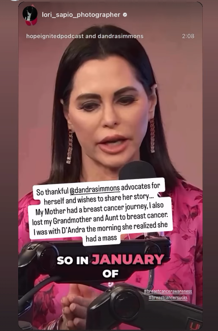
“In January of this year, I had gone in for an MRI, and I felt a mass on the right side of my breast; something didn’t feel right,” Simmons, 56, recalled in an Instagram story.

But it wasn’t a routine scan that brought the mass back to her attention—it was a weekend spent around barbecue smoke.
“You’re not going to believe this. This is all God,” Simmons said.
“I compete in barbecue, and I was at the Lone Star Smokeout as a guest. When I came home, I said, ‘I’m going to use this detox cream I have for my breasts because I’ve been around smoke all weekend.’ So, I started putting it on my breasts, and I said, ‘Wait,’ when I felt that mass on my breast.”
That moment of self-care turned into a life-saving realization. Simmons’ story is a powerful reminder of the importance of listening to your body—and staying proactive, especially when you’re at higher risk.
Simmons’ deep knowledge of her family history with cancer is something many people can model their thinking after for cancer prevention.
Expert Resources for Breast Cancer Screening
- 6 Common Excuses for Skipping a Mammogram That You Need to Stop Using!
- I Have Dense Breasts. Do I Need a 3D Mammogram?
- How to Avoid False Positive Cancer Results in Women With Dense Breasts: Ultrasounds Used in Addition To Mammograms
- The Mammogram Debate: Should Women Start Breast Cancer Screening at 30?
- When Should I Get a Mammogram?
- When You’re Called Back After a Mammogram: Breaking Down the Numbers
Family History & Breast Cancer Risk
Although breast cancer can happen to anyone, there are certain factors that can increase a person’s risk of getting the disease. The known risk factors for breast cancer include:
- Older age
- Having a gene mutation such as the BRCA1 or BRCA2
- Added exposure to estrogen
- Having children after the age of 30
- Exposure to radiation early in life
- Family history of the disease
Dr. Elizabeth Comen breaks down factors that may increase your breast cancer risk.
Some people think that breast cancer is only inherited through genes on the mother’s side, but it can also be related to a genetic mutation that can be found on the father’s side, according to breast oncologist Dr. Elizabeth Comen.
D’Andra’s Journey
Simmons, who appeared on RHOD from 2016 to 2021, publicly revealed her diagnosis in late July. Fortunately, her cancer was detected early—meaning the tumor was small and hadn’t spread to her lymph nodes—making it highly treatable.
Radiation was her latest treatment she received along her cancer journey.
This approach is commonly used after surgery to eliminate any remaining cancer cells in the breast or surrounding tissue. As Dr. Subhakar Mutyala explained to SurvivorNet, “Radiation therapy is actually ionizing energy… it causes DNA damage to destroy cancer cells.”
WATCH: Debates Around Radiation for Breast Cancer
Radiation treatment continues to evolve, with ongoing debates among experts about how to reduce side effects while optimizing outcomes. Dr. Chirag Shah, Director of Breast Radiation Oncology at Cleveland Clinic Cancer Center, outlined three key areas of discussion:
- Whole vs. Partial Breast Radiation: Shortening treatment duration and minimizing side effects are promising, though long-term data are still emerging.
- Identifying Patients Who May Not Need Radiation: Some individuals may not benefit from radiation, and omitting it could reduce unnecessary risks.
- Technique Optimization: Advancements in delivery methods aim to improve effectiveness while limiting harm to healthy tissue.
“I think the first debate that we have is whole breast radiation versus partial breast radiation and the idea of reducing duration of treatment and reducing side effects for patients, albeit with less than 10 years’ worth of long-term data,” Dr. Shah explained to SurvivorNet.
More on Radiation Therapy to Treat Breast Cancer
Radiation therapy—using high-energy rays to destroy cancer cells—is a common follow-up to breast cancer surgery, especially for patients who choose a lumpectomy over a mastectomy. Its goal: reduce the risk of recurrence by targeting any lingering cancer cells in the breast or surrounding tissue.
While effective, radiation can come with side effects, both immediate and delayed. Common symptoms include:
- Fatigue
- Swelling
- Scar tissue
- Shortness of breath
One of the more serious concerns involves the heart, which can unintentionally absorb radiation due to its proximity to the breast.
“When the radiation is delivered, unfortunately, the heart happens to be somewhere very near to where they have their breast cancer, and it becomes an innocent bystander absorbing some of the radiation,” explained Dr. Jean-Bernard Durand to SurvivorNet.
This exposure can lead to complications such as fatigue, shortness of breath, and even heart failure—sometimes surfacing decades after treatment.
“We make it a point to see them on a regular basis so that we can catch these things very early and treat them,” Dr. Durand added.
Even advanced techniques like proton therapy, which aim to minimize damage to healthy tissue, aren’t immune to side effects. Fatigue remains a common complaint, and the risk of long-term injury still exists.
“Radiation is a form of energy… and when we give radiation, it has the ability to scatter,” Dr. Durand said. “Even though we may target one particular area, that scattering of energy can cause injury to the local surrounding structures, including the heart.”
Over time, this injury can lead to the development of scar tissue within the heart muscle, its electrical system, and blood supply.
“We believe it is what causes all the injury, that ultimately leads to the symptoms,” he explained.
For survivors, this underscores the importance of ongoing monitoring and open conversations with care teams.
When to Screen for Breast Cancer
The medical community has a broad consensus that women should have annual mammograms between the ages of 45 and 54. However, an independent panel of experts called the U.S. Preventive Services Task Force (USPSTF) is saying that women should now start getting mammograms every other year at the age of 40, suggesting that this lowered age for breast cancer screening could save 19% more lives.
The American Cancer Society recommends getting a mammogram every other year for women 55 and older. However, women in this age group who want added reassurance can still get annual mammograms.
WATCH: When you’re getting a mammogram, ask about dense breasts.
Women with a strong family history of breast cancer, a genetic mutation known to increase the risk of breast cancer, such as a BRCA gene mutation, or a medical history, including chest radiation therapy before the age of 30, are considered at higher risk for breast cancer.
Experiencing menstruation at an early age (before 12) or having dense breasts can also put you into a high-risk category. If you are at a higher risk of developing breast cancer, you should begin screening earlier.
Screening Options for Women with Dense Breasts
Women with dense breasts should get additional screening to supplement their mammograms. Dense breasts mean more fibroglandular tissue, and less fatty breast tissue exists.
The dense tissue has a “masking effect on how well we can perceive cancer and find cancer on mammograms,” Dr. Cindy Ly, Chief of Breast Imaging and Vice Chair of Clinical Research and Faculty Affairs at the Stony Brook Renaissance School of Medicine, told SurvivorNet.
Glandular tissue within dense breasts appears white on mammograms, which can help mask potential cancer. The “frosted glass” effect from the glandular tissue can thus mask cancerous areas, especially developing ones. Undetected, these cancers can progress, growing large and advanced. They will then likely require more intensive treatments to cure, or can become incurable altogether.
WATCH: What is 3D Mammography?
“Digital mammography, it turns out, significantly improves the quality of the mammogram…It’s 3D or tomosynthesis mammography,” Dr. Connie Lehman, a diagnostic radiologist who specializes in breast cancer at Massachusetts General Hospital in Boston, explains.
“This allows us to find more cancers and to significantly reduce our false-positive rate. With digital mammography 3D tomosynthesis, we’re taking thin slices through that breast tissue, like slices of a loaf of bread. We can look at each slice independently rather than trying to see through the entire thickness of the loaf of bread. So those thin slices help us find things that were hidden in all the multiple layers,” Dr. Lehman adds.
Additional testing can be considered for dense breasts, depending on a woman’s personal history, preferences, and physician’s guidance. These tests include:
- 3-D Mammogram (Breast Tomosynthesis): This technology acquires breast imaging from multiple angles and digitally combines them into a 3D representation of the breast tissue. This allows physicians to see breast tissue architecture better, even in dense breasts. 3D mammograms are fast becoming the standard way of performing mammography.
- Breast Magnetic Resonance Imaging (MRI): An MRI machine uses magnets to create highly detailed, intricate images of the breast. These are mostly reserved for women with an extremely high breast cancer risk. Dense breasts alone may not be a valid reason to obtain a breast MRI. However, dense breasts in women with genetic mutations, like BRCA1 and BRCA2, or a strong family history of breast cancer, could justify obtaining breast MRIs.
- Molecular Breast Imaging (MBI): MBI is a newer imaging technique that uses a radioactive tracer to detect breast cancer. It is beneficial for women with dense breasts. However, MBI is not as widely available as other screening methods.
The Food and Drug Administration (FDA) now requires facilities offering mammograms to notify patients about their breast tissue density and recommend they speak with a doctor to determine if further screening is necessary. There will be “uniform guidance” on what language to use and what details to share with the patient to make the communication clear and understandable.
Understanding Your Mammogram Report
A radiologist reading mammograms categorizes breasts into four different categories using the Breast Imaging Reporting and Data System (BI-RADS), a classification system developed by the American College of Radiology (ACR). These include:
- Fatty breast tissue: These breasts are mainly fat with very little dense tissue. Found in less than 10% of women, fatty breasts appear dark on mammograms.
Scattered fibroglandular breast tissue: These breasts contain a mix of fatty and dense tissue (composed of glands and fibrous tissue). On a mammogram, they have dark areas (fatty tissue) intermixed with light areas (dense tissue). Around 40% of women have breasts that fall in this category. - Heterogeneously dense breast tissue: This type of breast tissue has many areas of dense tissue and some areas of fat. Found in 40% of women, these breasts look mostly light, with some dark areas on a mammogram.
- Extremely dense breast tissue: Such breasts are almost entirely composed of dense glandular and fibrous connective tissues with very little fat. They are found in 10% of women and appear light on mammograms.
Your breasts are usually called dense on a mammogram report if they fall within the heterogeneously dense breast tissue or the extremely dense breast tissue categories.
How Can I Manage Mammogram Anxiety?
It’s common to feel anxious while awaiting mammogram results. This feeling is often called mammogram anxiety.
If you are awaiting test results and your nerves are running rampant, try doing something that relaxes you, such as exercising or listening to your favorite music. Breathing exercises and meditation can also help you relax.
SurvivorNet has more resources to help you manage your mental health while awaiting test results.
Breast Cancer Symptoms & Self-Exams
Women are encouraged to do regular self-exams to become familiar with how their breasts feel normally, so when something unusual, like a lump, does form, it can be easily detected. A self-exam includes pressing your fingertips along your breast in a circular motion.
For some women, that means going to their doctor and walking through what a self-breast exam looks like, so they know what normal breast tissue feels like, so if they do feel something abnormal, whether it’s a lump or discharge from the nipple, they know what to ask and what to look for.
Below are common symptoms to look out for:
- New lump in the breast or underarm (armpit)
- Any change in the size or shape of the breast
- Swelling of all or part of the breast
- Skin dimpling or peeling
- Breast or nipple pain
- Nipple turning inward
- Redness or scaliness of the breast or nipple skin
- Nipple discharge (not associated with breastfeeding)
Learn more about SurvivorNet's rigorous medical review process.