
Mammograms, Ultrasounds & AI Are Tools to Help Detect Breast Cancer
- “The Office” actress Jenna Fischer’s early detection of Stage 1 triple‑positive breast cancer — discovered only after an initially inconclusive mammogram due to dense breast tissue underscores the lifesaving importance of consistent screening.
- Screening frequency remains a sticking point: the U.S. Preventive Services Task Force backs biennial mammograms starting at 40, while many experts argue annual screening catches more cancers earlier and saves more lives. If you have a higher risk for breast cancer due to a family history or a genetic mutation, you should consider screening at age 40.
- Enhanced mammography, including 3D mammography, “allows us to find more cancers and reduce false positives by viewing thin slices of breast tissue — like slicing a loaf of bread — to spot what’s hidden in the layers,” Dr. Connie Lehman, the chief of the Breast Imaging Division at Massachusetts General Hospital, explains.
- “AI‑based mammographic risk assessment creates an opportunity to identify women whose future risk may be higher even when traditional tools wouldn’t flag them,” says Dr. Beth Mittendorf from Dana‑Farber Cancer Institute and Chief of Breast Surgery at Beth Israel Deaconess Medical Center.
Fischer later explained that her dense breast tissue, which can make abnormalities harder to spot on imaging, likely contributed to the unclear results.

“Luckily my cancer was caught early, and it hadn’t spread,” she continued.
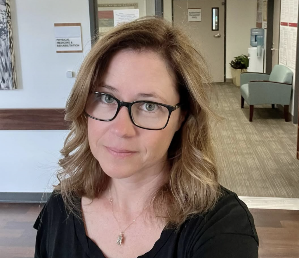
Fischer’s dense breasts meant it was more difficult to detect cancer on a regular mammogram. Because of this, some doctors turn to enhanced mammography.
Mammography saves lives — that part isn’t up for debate. But how often women should be screened, and how dense breast tissue factors into those decisions, remains a point of tension.
Mammography’s benefit in reducing breast cancer deaths is widely accepted, yet concerns about overdiagnosis keep screening guidelines under scrutiny. The U.S. Preventive Services Task Force recommends biennial screening for women 40–74, noting a moderate net benefit and emphasizing equity, especially because Black women face a 40% higher breast‑cancer mortality rate than White women.
Critics argue that annual screening catches more late‑stage cancers and saves more years of life. Professional groups like the American College of Obstetricians and Gynecologists advise average‑risk women to screen every one to two years starting at 40, while radiology organizations often favor yearly screening.
Dense breasts add another layer of complexity. Dense tissue appears white on a mammogram — the same color as many cancers — making tumors easier to miss and modestly increasing risk. Since September 2024, the FDA now requires all U.S. mammography reports to include breast‑density assessments, ensuring patients know whether they fall into the heterogeneously dense or extremely dense categories.
WATCH: When Should I Get a Mammogram?
Dr. Connie Lehman, a diagnostic radiologist who specializes in breast cancer at Massachusetts General Hospital in Boston, has advice for women.
“If you haven’t gone through menopause yet, I think it’s very important that you have a mammogram every year. We know that cancers grow more rapidly in our younger patients, and having that annual mammogram can be lifesaving,” Dr. Lehman tells SurvivorNet.
“After menopause, it may be perfectly acceptable to reduce that frequency to every two years,” Dr. Lehman continued.
AI is also helping mammography with Clairity Breast, an FDA-cleared artificial intelligence program that analyzes subtle patterns in screening mammograms; it doesn’t detect cancer in real time. Instead, it predicts a woman’s future breast cancer risk, generating a validated five‑year risk score.
The NCCN guidelines include an AI-based mammogram risk assessment option. The update recommends that when AI identifies a patient as high risk, clinicians take direct action, such as offering supplemental imaging or discussing risk-reduction strategies.

“Historically, breast cancer risk assessment has relied on age, family history, genetics, and breast density,” explains Dr. Beth Mittendorf, Chief of Multidisciplinary Oncology at Dana‑Farber Cancer Institute and Chief of Breast Surgery at Beth Israel Deaconess Medical Center.
“AI‑based mammographic risk assessment creates an opportunity to identify women whose future risk may be higher even when traditional tools wouldn’t flag them,” Dr. Mittendorf adds.
“This new approach can enhance the current standard of care,” says Dr. Mary Newell, breast imaging specialist at Winship Cancer Institute of Emory University.
Dr. Basak Dogan, Director of Breast Imaging Research at UT Southwestern’s Simmons Cancer Center, notes that AI may reclassify many women previously considered average risk.
“Patients without a strong family history may now be identified as at increased risk based on their specific breast tissue patterns,” she says.
“That means more women could qualify for supplemental screening earlier—potentially catching cancers that would otherwise be missed,” Dr. Dogan continued.
Helpful Information on Mammograms
- 6 Common Excuses for Skipping a Mammogram That You Need to Stop Using!
- I Have Dense Breasts. Do I Need a 3D Mammogram?
- Mammograms Are Still the Best Tool for Detecting Breast Cancer — A Warning About Thermography
- New Guidelines Say Many Women Under 50 Can Skip Mammograms. That May Not Be The Best Advice
- Successful Doctor’s Breast Cancer Missed On A Mammogram. Asymmetrical Breasts Lead to Diagnosis
What To Know About Enhanced Mammograms
Enhanced mammograms are a helpful tool for women whose breast tissues obscure precise mammography readings. While implants often call for enhanced mammogram screening, women with dense breasts may also need enhanced screening. Dense breasts mean more fibroglandular tissue and less fatty breast tissue.
During mammograms, the tissue difference can make locating signs of cancer more difficult in women with dense breasts.
Additional testing can be considered for these women, depending on their personal history, preferences, and their physician’s guidance.
These tests include:
- 3-D Mammogram (Breast Tomosynthesis): This technology acquires breast imaging from multiple angles and digitally combines them into a 3D representation of the breast tissue. This allows physicians to see breast tissue architecture better, even in dense breasts. 3D mammograms are fast becoming the standard way of performing mammography.
- Breast Magnetic Resonance Imaging (MRI): An MRI machine uses magnets to create highly detailed, intricate images of the breast. These are mostly reserved for women with an extremely high breast cancer risk. Dense breasts alone may not be a valid reason to obtain a breast MRI. However, dense breasts in women with genetic mutations, like BRCA1 and BRCA2, or a strong family history of breast cancer, could justify obtaining breast MRIs.
- Molecular Breast Imaging (MBI): MBI is a newer imaging technique that uses a radioactive tracer to detect breast cancer. It is beneficial for women with dense breasts. However, MBI is not as widely available as other screening methods.
When you’re getting a mammogram, ask about dense breasts.
Understanding Your Mammogram Report
A radiologist reading mammograms categorizes breasts into four different categories using the Breast Imaging Reporting and Data System (BI-RADS), a classification system developed by the American College of Radiology (ACR).
These include:
- Fatty breast tissue: These breasts are mainly fat with very little dense tissue. Found in less than 10% of women, fatty breasts appear dark on mammograms.
- Scattered fibroglandular breast tissue: These breasts contain a mix of fatty and dense tissue (composed of glands and fibrous tissue). On a mammogram, they have dark areas (fatty tissue) intermixed with light areas (dense tissue). Around 40% of women have breasts that fall in this category.
- Heterogeneously dense breast tissue: This type of breast tissue has many areas of dense tissue and some areas of fat. Found in 40% of women, these breasts look mostly light, with some dark areas on a mammogram.
- Extremely dense breast tissue: Such breasts are almost entirely composed of dense glandular and fibrous connective tissues with very little fat. They are found in 10% of women and appear light on mammograms.
Your breasts are usually called dense on a mammogram report if they fall within the heterogeneously dense breast tissue or the extremely dense breast tissue categories.
When To Screen For Breast Cancer
Recently, an independent panel of experts called the U.S. Preventive Services Task Force (USPSTF) upgraded its guidelines to suggest that women start getting mammograms every other year at the age of 40. The task force notes that this lowered the age (the previous recommendation was to begin screening at age 45) for breast cancer screening could save 19% more lives.
The American Cancer Society recommends getting a mammogram every other year for women 55 and older. However, women in this age group who want added reassurance can still get annual mammograms.
Mammograms are still the best tool for detecting breast cancer.
Women with a strong family history of breast cancer, a genetic mutation known to increase the risk of breast cancer, such as a BRCA gene mutation, or a medical history, including chest radiation therapy before the age of 30, are considered at higher risk for breast cancer.
Experiencing menstruation at an early age (before 12) or having dense breasts can also put you into a high-risk category. If you are at a higher risk of developing breast cancer, you should begin screening earlier.
More on Fischer’s Cancer Journey and Road to Remission
“In January [2024], I had a lumpectomy to remove the tumor,” Fischer explained. A lumpectomy is a procedure that removes the tumor and some of the surrounding tissue.
“Luckily, my cancer was caught early, and it hadn’t spread into my lymph nodes or throughout the rest of my body; however, because of the aggressive nature of triple-positive breast cancer, it still required chemotherapy and radiation to be sure it didn’t return,” Fischer added.
She underwent 12 rounds of weekly chemotherapy in February 2024 and began three weeks of radiation in June 2024.

“I lost my hair during chemotherapy, but thanks to some great wigs and hats with hair, I have been able to wait until now to reveal all that has been going on for me.”
“In January [2024], I had a lumpectomy to remove the tumor,” Fischer explained. A lumpectomy is a procedure that removes the tumor and some of the surrounding tissue.
“Luckily, my cancer was caught early, and it hadn’t spread into my lymph nodes or throughout the rest of my body; however, because of the aggressive nature of triple-positive breast cancer, it still required chemotherapy and radiation to be sure it didn’t return,” Fischer added.

She underwent 12 rounds of weekly chemotherapy in February 2024 and began three weeks of radiation in June 2024.
“In January [2024], I had a lumpectomy to remove the tumor,” Fischer explained. A lumpectomy is a procedure that removes the tumor and some of the surrounding tissue.
“Luckily, my cancer was caught early, and it hadn’t spread into my lymph nodes or throughout the rest of my body; however, because of the aggressive nature of triple-positive breast cancer, it still required chemotherapy and radiation to be sure it didn’t return,” Fischer added.
She underwent 12 rounds of weekly chemotherapy in February 2024 and began three weeks of radiation in June 2024.
“I lost my hair during chemotherapy, but thanks to some great wigs and hats with hair, I have been able to wait until now to reveal all that has been going on for me,”
Hair loss is challenging for women and men alike, but it can be incredibly difficult for cancer patients. Losing your hair or seeing it thinning is often a side effect of some cancer treatments.
“For cancer patients, losing one’s hair can be unbelievably stressful. To start with, the dread of losing one’s hair can lead to some sleepless nights and feelings of anxiety,” Dr. Samantha Boardman, a New York-based psychiatrist and author, told SurvivorNet.
To cope, Dr. Boardman suggested reaching out to other survivors who have been through a similar situation.
Chemotherapy can cause hair loss. It usually begins about three to four weeks after starting chemotherapy and continues throughout treatment.
This happens because this treatment targets quickly dividing cells throughout the body, including cancer cells and hair cells.
Radiation is another treatment that can lead to hair loss if the hair is in the path of the tumor being treated. Radiation for a brain tumor, for example, may cause hair loss.
“If you do lose hair, it will regrow several weeks or months after treatment,” radiation oncologist Dr. James Taylor told SurvivorNet. “Fortunately, for most patients, hair loss is not a concern when having radiation therapy.”
Fortunately, hair loss during cancer treatment is not all bad news. Most people can expect regrowth four to six weeks after treatment. However, when your hair grows back, you may notice some changes in its color and texture.
If losing your hair is a concern for you before cancer treatment, know you have options like wigs, hats, and wraps, as Fischer used.

Although Fischer had Shaerer to help her through the ups and downs of her cancer journey, Fischer took to Instagram to encourage others battling the disease.
“I’m also sharing in hopes that it will be a source of support to any woman who is going through this right now,” Fischer said.
“Suddenly, everything in your life is geared around one thing: fighting cancer,” Fischer said.
Fischer revealed a year after her diagnosis that she had reached remission.
“After completing surgery, chemotherapy, and radiation, I am now cancer-free,” Fischer said.
Radiation Therapy: A Powerful Tool with Long-Term Considerations
Fischer’s treatments included radiation therapy and chemotherapy.
Radiation therapy—using high-energy rays to destroy cancer cells—is a common follow-up to breast cancer surgery, especially for patients who choose a lumpectomy over a mastectomy. Its goal: reduce the risk of recurrence by targeting any lingering cancer cells in the breast or surrounding tissue.
While effective, radiation can come with side effects, both immediate and delayed. Common symptoms include:
- Fatigue
- Swelling
- Scar tissue
- Shortness of breath
One of the more serious concerns involves the heart, which can unintentionally absorb radiation due to its proximity to the breast.
“When the radiation is delivered, unfortunately, the heart happens to be somewhere very near to where they have their breast cancer, and it becomes an innocent bystander absorbing some of the radiation,” explained Dr. Jean-Bernard Durand to SurvivorNet.
This exposure can lead to complications such as fatigue, shortness of breath, and even heart failure—sometimes surfacing decades after treatment.
“We make it a point to see them on a regular basis so that we can catch these things very early and treat them,” Dr. Durand added.
Even advanced techniques like proton therapy, which aim to minimize damage to healthy tissue, aren’t immune to side effects. Fatigue remains a common complaint, and the risk of long-term injury still exists.
“Radiation is a form of energy… and when we give radiation, it has the ability to scatter,” Dr. Durand said. “Even though we may target one particular area, that scattering of energy can cause injury to the local surrounding structures, including the heart.”
Over time, this injury can lead to the development of scar tissue within the heart muscle, its electrical system, and blood supply.
“We believe it is what causes all the injury, that ultimately leads to the symptoms,” he explained.
For survivors, this underscores the importance of ongoing monitoring and open conversations with care teams.
Radiation treatment continues to evolve, with ongoing debates among experts about how to reduce side effects while optimizing outcomes. Dr. Shah outlined three key areas of discussion:
- Whole vs. Partial Breast Radiation: Shortening treatment duration and minimizing side effects are promising, though long-term data are still emerging.
- Identifying Patients Who May Not Need Radiation: Some individuals may not benefit from radiation, and omitting it could reduce unnecessary risks.
- Technique Optimization: Advancements in delivery methods aim to improve effectiveness while limiting harm to healthy tissue.
“I think the first debate that we have is whole breast radiation versus partial breast radiation and the idea of reducing duration of treatment and reducing side effects for patients, albeit with less than 10 years’ worth of long-term data,” Dr. Shah explained to SurvivorNet.
Managing Chemotherapy and Its Side Effects
Chemotherapy is an effective tool for oncologists to help treat cancer by stopping cancerous cells from growing, dividing, and spreading to other organs. Chemo works by traveling through the bloodstream, killing cancerous cells. However, healthy cells are also impacted in the process, leading to side effects.
Patients almost universally experience fatigue, often alongside gastrointestinal side effects, such as nausea. Doctors have many effective medications to combat chemo-induced nausea. “But mitigating that fatigue often depends on the patient,” says Dr. Renata Urban, a gynecologic oncologist at the University of Washington in Seattle.
“Neuropathy is probably one of the most challenging side effects,” says Dr. Renata Urban, a gynecologic oncologist at the University of Washington in Seattle. Neuropathy results from damage to the peripheral nerves. It usually resolves after chemotherapy treatment, but sometimes symptoms can persist. While it’s typically characterized by numbness or a pins-and-needles sensation in the hands and feet, neuropathy can have several different symptoms, including:
- Weakness in the hands or feet
- Stabbing or burning pain in the hands or feet
- Difficulty gripping, such as when holding a fork
- Difficulty with fine motor skills, such as writing or buttoning a shirt
Nausea and vomiting are common side effects of chemotherapy. When chemotherapy affects the rapidly dividing cells in the lining of the stomach, the resulting cellular havoc in the gastrointestinal tract can lead to side effects such as nausea and vomiting. However, doctors can help patients mitigate the hit with various medications before, during, and after treatment.
“Part of the chemotherapy prescription includes a set regimen of anti-nausea medications,” says Dr. Renata Urban, a gynecologic oncologist at the University of Washington in Seattle. “We also ensure that patients have medications at home that they can use should they develop nausea after treatment.”
Hair loss is another side effect of chemotherapy.
WATCH: Coping with hair loss.
“For cancer patients, losing one’s hair can be unbelievably stressful. To start with, the dread of losing one’s hair can lead to some sleepless nights and feelings of anxiety,” Dr. Samantha Boardman, a New York-based psychiatrist and author, told SurvivorNet.
Chemotherapy can cause hair loss. It usually begins about three to four weeks after chemotherapy and continues throughout treatment.
It happens because this treatment targets quickly dividing cells throughout the body. That includes cancer cells but also hair cells.
Most patients can expect regrowth four to six weeks after treatment. However, it is possible that when your hair grows back, you may notice some changes in its color and texture.
RELATED: How are chemotherapy side effects managed for ovarian cancer treatment?
Tips for Navigating Chemo Side Effects
When dealing with fatigue, doctors don’t have an arsenal of weapons to combat fatigue in terms of prescription medications. However, you can do several things to help minimize the hit and restore your energy.
- Exercise: While it may be counterintuitive, physical activity can help alleviate side effects, especially fatigue. “Although ovarian cancer is not common, we often draw upon the experience of patients with breast cancer and colon cancer, who have shown that physical activity can not only improve quality of life but may also have beneficial impacts on cancer outcomes,” Dr. Urban says.
- Eat well: Even though nausea may interfere with your ability to eat a healthy diet, it’s essential to ensure you’re eating appropriately, getting enough protein, and not losing weight. Not only will nourishing your body support your recovery, but it may also help you feel more energized.
- Sleep: Want to mitigate fatigue? Be sure to maintain your regular sleep-wake cycle while on treatment. Sticking to a set sleep schedule helps reduce fatigue by ensuring enough hours for your body to heal and restore itself each night. It may also help you recover more quickly by keeping energy levels high during the daytime.
Treating Neuropathy Symptoms
Doctors have several strategies for helping patients deal with this side effect. Once a patient begins experiencing the symptoms of neuropathy, they’ll be carefully monitored to make sure it doesn’t get worse. Before each chemotherapy infusion, the attending oncologist will assess whether the symptoms have progressed. If the symptoms worsen, they may adjust the dose or delay treatment. They may also try switching to another chemotherapy drug.
How to Get a Handle on Nausea
Most of these anti-nausea medications last for more than eight hours. One of the infusions commonly used reduces the degree of nausea for up to three days.
Complementary approaches may also be helpful. A few favorites:
- Ginger: Studies consistently show that ginger helps alleviate chemotherapy-induced nausea. The powerful herb appears to have an anti-spasmodic effect on the gut. Not a fan of raw ginger? Suck on ginger candy, sip ginger ale, or make a steaming cup of ginger tea.
- Pressure bracelets: at your local pharmacy, these bracelets provide consistent pressure on a particular acupressure point on the wrist to reduce nausea.
- Deep breathing: Moving air in and out of your lungs with a few deep breaths can help relieve nausea, particularly if you pair deep breathing exercises with meditation. It can also help you relax and release stress and anxiety.
WATCH: Managing chemo side effects.
Coping with Hair Loss
If losing your hair is a concern for you before cancer treatment, know you have options like wigs, hats, wraps, and scarves, among other things.
Another option that can minimize hair loss is cryotherapy, “just a fancy way for saying cold therapy,” says Dr. Renata Urban, gynecologic oncologist at the University of Washington in Seattle.
Cryotherapy involves wearing cold caps or special cooling caps before, during, and after each chemotherapy treatment.
What To Ask Your Doctor
If you have been diagnosed with breast cancer, you may have questions about keeping your strength through treatment. Here are a few questions to help you begin the conversation with your doctor:
- What treatment will I be receiving?
- What side effects are associated with this treatment?
- Are there steps I can take daily to help minimize these side effects?
- What physical activity routine do you recommend for me during treatment?
- Do you have recommendations for someone who doesn’t particularly enjoy exercise?
- Can you recommend a dietician who can help me with healthy eating tips and weight maintenance?
- I’ve been having trouble sleeping. Do you have any treatment recommendations?
Learn more about SurvivorNet's rigorous medical review process.


