
The Importance of Breast Cancer Screenings
- Former Good Morning America host Joan Lunden, 76, says her annual mammogram came back clear, 12 years after her triple-negative breast cancer diagnosis.
- Lunden, who has dense breasts, also gets an ultrasound every year. In 2014, her mammogram nearly missed her cancer. She has urged postmenopausal women to add a bone density test to their annual screenings.
- Current federal guidance recommends that women at average risk get a mammogram every other year from age 40 to 74; some groups, including the American Cancer Society, recommend yearly screening for certain ages.
- Women at high risk because of a genetic mutation such as BRCA1 or BRCA2 or other factors should talk with their doctor about starting earlier
- Nearly half of women have dense breasts, which both raises breast cancer risk and can hide tumors on a mammogram. 3D mammography, along with supplemental tests such as breast ultrasound, MRI and molecular breast imaging, may help, so ask your doctor about your breast density and your options.
“Started my day with my annual mammogram, and I’m happy to report it came back clear!” Lunden wrote on Instagram, posting a video filmed inside the radiology office where she gets screened every year.
Read More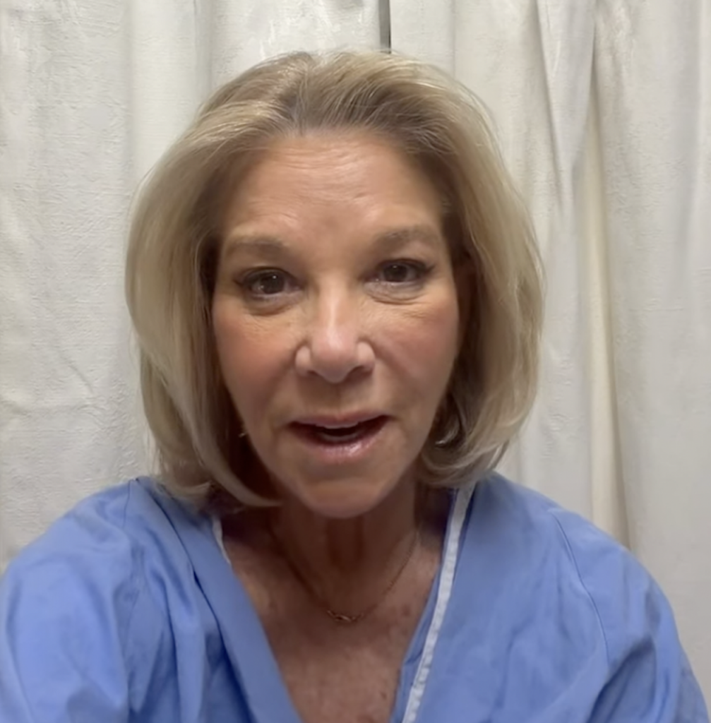
Expert Resources on Breast Cancer Screening
- Bi-Annual Mammograms At Age 40 Now Recommended For Most Women, What The New Breast Cancer Screening Guidelines Mean For You
- Black Women May Need To Start Breast Cancer Screening At 42, According To A New Study
- Breast Cancer Screening: When to Start, and Why Your Risk Score Matters
- Breast Cancer: Introduction to Prevention & Screening
- Doctors Say New FDA-Approved Breast Cancer Screening Tool Using AI Won’t Replace Traditional Mammograms But Will Enhance Them
- The Mammogram Debate: Should Women Start Breast Cancer Screening at 30?
- How to Avoid False Positive Cancer Results in Women With Dense Breasts: Ultrasounds Used in Addition To Mammograms
Then, without missing a beat, she turned her good news into a public-service message, telling her fans, “I added a bone density test today and if you are postmenopausal, make sure you add that as well.”
“So make sure you have your annual screenings every year,” concluded Lunden, who looked healthy and relaxed in the footage, with her signature honey-blonde bob swept to the side in the polished style fans remember from her GMA years.
Triple-negative breast cancer, the type Lunden battled, lacks three key biomarkers: the estrogen receptor (ER), the progesterone receptor (PR) and HER2. That makes it more difficult to treat, because hormone therapies and HER2-targeted drugs don’t work against it.
“Any triple-negative cancer that’s over half a centimeter or has lymph node involvement needs chemotherapy,” medical oncologist Dr. Julie Nangia previously told SurvivorNet.
Treatment typically combines two classes of chemotherapy: taxanes, such as paclitaxel (Taxol), which stop cancer cells from dividing, and anthracyclines, such as doxorubicin (Adriamycin), which damage cancer cells’ DNA. They are often given before surgery to shrink the tumor. For many patients with higher-risk early-stage disease, the immunotherapy drug pembrolizumab (Keytruda) is now added before and after surgery.
Joan Lunden’s Cancer Journey
For 17 years, from 1980 to 1997, Lunden was one of the most familiar faces in American mornings as co-host of ABC’s Good Morning America. She later moved to NBC’s TODAY as a special correspondent, and it was during that run that she learned she had cancer in her right breast. Her choice to go through that diagnosis in public has since become as central to her legacy as anything she did on air.
Lunden’s insistence on adding an ultrasound is personal. She was 63 when she was diagnosed with stage two triple-negative breast cancer in 2014 — and her annual mammogram that year nearly missed it. Her doctors initially saw nothing significant, because Lunden has dense breasts.
“Candidly, I never thought I would be one of the women who would get breast cancer,” Lunden told SurvivorNet in an earlier interview.
WATCH: Joan Lunden Ages With Candor
Dense breasts contain less fatty tissue, which makes it harder for a mammogram alone to spot cancer, so women with dense breasts are encouraged to add an ultrasound.
Dr. Linda Pak, a breast cancer surgeon/oncologist at NYU Langone’s Perlmutter Cancer Center and clinical assistant professor of surgery at NYU Grossman School of Medicine, explained to SurvivorNet:
“Ultrasound screening is a very helpful supplemental method of imaging, to be used in addition to a mammogram, for women with dense breasts. Increased breast density can make tumors more difficult to see on mammograms. Ultrasound can detect additional cancers that are not seen on mammography.
“Therefore I recommend a supplemental screening breast ultrasound in addition to mammography for all patients with dense breasts.”
In her memoir, “Had I Known,” Lunden wrote of her surprise that dense tissue had made her tumor hard to detect.
WATCH: Joan Lunden Helps Best-selling author Laura Mortons Through Cancer
Helpful Information About Breast Cancer Screening
The American Cancer Society recommends annual mammograms for women ages 45 to 54, with the option to start yearly screening at 40. For women 55 and older, it recommends a mammogram every other year, though women who want added reassurance can continue annual screening.
The U.S. Preventive Services Task Force (USPSTF), an independent panel of experts, updated its guidance in 2024. It now recommends that all women at average risk get a mammogram every other year from age 40 through 74, estimating that starting screening at 40 instead of 50 would save more lives.
RELATED: ‘Annual’ Screenings For Breast Cancer Survivors May Not Be Necessary, New Study Suggests
Women are considered at high risk for breast cancer if they have a strong family history of the disease, a genetic mutation known to raise breast cancer risk (such as BRCA1 or BRCA2), or a history of chest radiation therapy before age 30. Women at high risk should talk with their doctor about starting screening earlier and about whether they need additional tests, such as breast MRI.
WATCH: Understanding the BRCA gene mutation
Other factors, such as starting menstruation early (before age 12) or having dense breasts, also raise breast cancer risk, though usually not enough on their own to put a woman in the high-risk category.
Breast density is determined through mammograms, and mammogram reports are now required to tell patients whether they have dense breasts. Dense breast tissue raises a woman’s risk of breast cancer, and it can also hide tumors on a mammogram. 3D mammography can improve detection, and supplemental tests such as breast ultrasound, breast MRI and molecular breast imaging may also be options for women with dense breasts. It is important to ask your doctor about your breast density and cancer risk.
RELATED: 3D Mammography Detects 34% More Breast Cancers Than Traditional Mammography
Family History & Breast Cancer Risk
Although breast cancer can happen to anyone, certain factors can increase a person’s risk of getting the disease. The known risk factors for breast cancer include:
- Older age
- Having a gene mutation such as BRCA1 or BRCA2
- Added exposure to estrogen
- Having a first child after age 30, or never having children
- Exposure to radiation early in life
- Family history of the disease
RELATED: Is Genetic Testing Right for You?
About ten percent of breast cancers are hereditary, says Dr. Ophira Ginsburg, Director of the High-Risk Cancer Program at NYU Langone’s Perlmutter Cancer Center.
“We encourage only those with a family history to get [genetic testing],” Dr. Ginsburg previously told SurvivorNet. “I would say that if you have anyone in your family diagnosed with a rare cancer. Or if you have a strong family history of one or two kinds of cancer, particularly breast and ovarian.”
She noted that a family history of colon, rectal and uterine cancers can also point to an inherited condition called Lynch syndrome.
There are two main types of genetic testing. The first, hereditary genetic testing, helps people with a family history of cancer better understand their own risk. It is usually done with a blood or saliva test, and your doctor will discuss your family history in the context of your tumor type and your age at diagnosis.
WATCH: Understanding genetic testing for breast cancer.
The second involves genetic sequencing of your tumor if you’ve been diagnosed with cancer. These genetic changes can be inherited, but most arise during a person’s lifetime. This process usually involves examining a biopsy or surgical specimen of your tumor, and it can lead to decisions on drugs that might work against your cancer.
“Digital mammography, it turns out, significantly improves the quality of the mammogram… It’s 3D or tomosynthesis mammography,” Dr. Connie Lehman, the chief of the Breast Imaging Division at Massachusetts General Hospital, told SurvivorNet in an earlier interview.
“This allows us to find more cancers and significantly reduce our false-positive rate. With digital mammography 3D tomosynthesis, we’re taking thin slices through that breast tissue, like slices of a loaf of bread. We can look at each slice independently rather than trying to see through the entire thickness of the entire loaf of bread. So those thin slices help us find things that were hidden in all the multiple layers,” Dr. Lehman adds.
Additional testing can be considered for dense breasts, depending on a woman’s personal history, preferences, and her physician’s guidance.
Understanding Your Mammogram Report and Breast Density
A radiologist reading mammograms categorizes breasts into four different categories using the Breast Imaging Reporting and Data System (BI-RADS), a classification system developed by the American College of Radiology (ACR). These include:
- Fatty breast tissue: These breasts are mainly composed of fat with very little dense tissue. Found in less than 10% of women, fatty breasts appear dark on mammograms.
- Scattered fibroglandular breast tissue: These breasts contain a mix of fatty and dense tissue (composed of glands and fibrous tissue). On a mammogram, they have dark areas (fatty tissue) intermixed with light areas (dense tissue). Around 40% of women have breasts that fall in this category.
- Heterogeneously dense breast tissue: This type of breast tissue has many areas of dense tissue and some areas of fat. Found in 40% of women, these breasts look mostly light, with some dark areas on a mammogram.
- Extremely dense breast tissue: Such breasts are almost entirely composed of dense glandular and fibrous connective tissues with very little fat. They are found in 10% of women and appear light on mammograms.
Your breasts are usually called dense on a mammogram report if they fall within the heterogeneously dense breast tissue or the extremely dense breast tissue categories.
WATCH: 3D Mammograms explained.
Additional testing includes:
- 3-D Mammogram (Breast Tomosynthesis): This technology acquires breast imaging from multiple angles and digitally combines them into a 3D representation of the breast tissue. This allows physicians to see breast tissue architecture better, even in dense breasts. 3D mammograms are fast becoming the standard way of performing mammography.
- Breast Magnetic Resonance Imaging (MRI): An MRI machine uses magnets to create highly detailed, intricate images of the breast. These are mostly reserved for women with an extremely high breast cancer risk. Dense breasts alone may not be a valid reason to obtain a breast MRI. However, dense breasts in women with genetic mutations, like BRCA1 and BRCA2, or a strong family history of breast cancer, could justify obtaining breast MRIs.
- Molecular Breast Imaging (MBI): MBI is a newer imaging technique that uses a radioactive tracer to detect breast cancer. It is beneficial for women with dense breasts. However, MBI is not as widely available as other screening methods.
Since September 2024, a Food and Drug Administration (FDA) rule has required facilities offering mammograms to notify patients about their breast tissue density, using standard language, and to recommend that they speak with a doctor about whether further screening is necessary.
Breast Density Doesn’t Remain the Same Over Time, Impacting Your Cancer Risk
Women with dense breasts are at a higher risk of developing breast cancer, a connection shown in several rigorous scientific studies. The exact reason for this difference is not fully understood.
In a 2024 study of 947 women by researchers at Washington University School of Medicine in St. Louis and Brigham and Women’s Hospital in Boston, breast density declined with age, but women who went on to develop breast cancer showed a slower decline than those who did not. The researchers concluded that how quickly density changes may help signal future breast cancer risk.
WATCH: Mammograms are still the best tool for detecting breast cancer.
Regular Self-Exams Are Helpful In Between Mammograms
Knowing how your breasts normally look and feel can help you spot changes between mammograms. Watch for lumps, swelling, bulging, or changes in the shape of the breast or nipple.
Checking for signs of redness, rashes, or nipple discharge is also important. If you notice anything concerning, contact your doctor. Being familiar with your breasts is a complement to regular mammograms, not a replacement.
WATCH: How to perform a self-exam.
How AI Is Enhancing Mammograms When Screening For Breast Cancer
An artificial intelligence program authorized by the Food and Drug Administration (FDA), called Clairity Breast, may change the way doctors assess breast cancer risk from screening mammograms.
While not a replacement for traditional mammography, this AI-powered tool can enhance current screening practices and improve early detection efforts, according to Dr. Mary Newell, a breast imaging specialist at Winship Cancer Institute of Emory University.
“This new approach can enhance the current standard of care,” Dr. Newell tells SurvivorNet.
Clairity Breast analyzes subtle patterns in screening mammograms linked to future breast cancer risk, generating a validated five-year risk score. This score is integrated into clinical systems, enabling more personalized follow-up care.
“We can identify patients who are at elevated risk and offer supplemental screening to them with breast MRI or other technologies as a way to increase the likelihood of early detection. It does not replace mammography, and in fact, relies on mammographic images to allow the risk assessment,” she adds.
Currently, women at average risk are advised to begin regular mammograms at age 40. While Clairity Breast does not change these guidelines, Dr. Newell noted that it could help identify patients who may need earlier screening or additional imaging methods.
“It may allow us to identify patients who should start screening at an earlier age if they are shown to be at elevated risk,” she said. “It can also allow us to identify patients who may benefit from supplemental screening technologies, in addition to their yearly mammogram, after the age of 40.”
Unlike AI programs designed to detect cancer directly from images, Clairity Breast functions as a risk assessment tool. It does not interpret mammograms for cancer detection but instead works alongside traditional methods to pinpoint patients who may be at higher risk.
“Other AI tools are being developed that provide assistance with interpretation, helping to detect a cancer that may already be present,” Dr. Newell explained. “But most of the literature to date suggests that AI tools work best when deployed in conjunction with interpretation by a breast radiologist.”
Clairity Breast represents a promising step in breast cancer prevention, offering a new layer of analysis that helps doctors identify high-risk patients early and refine screening approaches, all while keeping mammography at the core of detection.
“Now, advancements in AI and computer vision can uncover hidden clues in mammograms — invisible to the human eye — to help predict future risk,” said Dr. Lehman, founder of Clairity and breast imaging specialist at Mass General Brigham, in a press release.
Dr. Amani Jambhekar, a board-certified surgeon specializing in breast cancer and melanoma at CHRISTUS Health, says this new technology can make mammograms even more valuable.
“It may be a more individualized way of calculating breast cancer risk, which we need, as existing risk calculators do not identify everyone who is high risk. As a surgical oncologist, I am excited to learn more about this platform when it launches,” Dr. Jambhekar tells SurvivorNet.
How is 3D mammography different?
False Positives & the Dreaded ‘Callback’
Getting a call from the doctor’s office asking you to come back for more images, such as an MRI or ultrasound, is worrying, but it can mean a number of things. Often the initial pictures simply weren’t clear. For instance, dense breasts can make it hard to distinguish between fatty tissue and a tumor.
Here is how the numbers on screenings break down, according to Dr. Lehman: Of every 1,000 women screened, about 100 are called back, and the vast majority will be absolutely fine after more imaging. About 20 will need a biopsy, and in the end, about 5 will be found to have cancer.
Here are some tips to help ensure you get the most accurate test possible:
- Go to a large and experienced center that has a good record and reputation.
- Don’t wear deodorant on the day of your mammography. It can contribute to inaccuracies.
- Keep in mind that certain factors, like dense breasts, tattoos, and breast implants, may also result in errors.
Contributing: SurvivorNet Staff
Learn more about SurvivorNet's rigorous medical review process.


